CytoFix™ Red Lysosomal Stain
Lysosomes are cellular organelles which contain acid hydrolase enzymes to break up waste and cellular debris through a process known as autophagy. AAT Bioquest offers CytoFix™ Red lysosomal stain for selectively staining lysosomes. CytoFix™ Red lysosomal stain is well retained in lysosomes even after fixation. The dye permeates intact live cells and gets trapped in lysosomes. The fluorescence in lysosomes generated by this dye is well retained at least for 1 week, making it an excellent lysosomal tracking dye. The key features of this stain are its high staining efficiency, long retention after fixation with minimal hands on time. CytoFix™ Red lysosomal stain can be used with GFP expressed cells or with other organelles stains for multicolor analysis. It can be used for both suspension and adherent cells and readily adapted for a wide variety of fluorescence platforms.
Platform
Fluorescence microscope
| Excitation | Cy3/TRITC filter set |
| Emission | Cy3/TRITC filter set |
| Recommended plate | Black wall/clear bottom |
| Instrument specification(s) | Cy3/TRITC filter set |
Calculators
Common stock solution preparation
Table 1. Volume of DMSO needed to reconstitute specific mass of CytoFix™ Red Lysosomal Stain to given concentration. Note that volume is only for preparing stock solution. Refer to sample experimental protocol for appropriate experimental/physiological buffers.
| 0.1 mg | 0.5 mg | 1 mg | 5 mg | 10 mg | |
| 1 mM | 115.774 µL | 578.871 µL | 1.158 mL | 5.789 mL | 11.577 mL |
| 5 mM | 23.155 µL | 115.774 µL | 231.548 µL | 1.158 mL | 2.315 mL |
| 10 mM | 11.577 µL | 57.887 µL | 115.774 µL | 578.871 µL | 1.158 mL |
Molarity calculator
Enter any two values (mass, volume, concentration) to calculate the third.
| Mass (Calculate) | Molecular weight | Volume (Calculate) | Concentration (Calculate) | Moles | ||||
| fgpgngµgmgg | / | g/mol | = | fLpLnLµLmLL | x | fMpMnMµMmMM | = | fmolpmolnmolµmolmmolmol |
Spectrum
Open in Advanced Spectrum Viewer
Spectral properties
Images
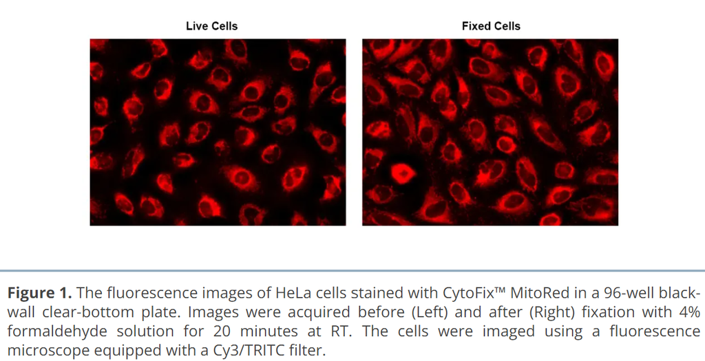
 Figure 1. The fluorescence images of HeLa cells stained with CytoFix™ LysoRed in a 96-well black-wall clear-bottom plate. Image was acquired before (Left) and after (Right) fixation with 4% formaldehyde solution for 20 minutes at RT. The cells were imaged using fluorescence microscope with a Cy3/TRITC filter.
Documents
Safety data sheet
Product protocol
Certificate of analysis
Application notes
A Novel Fluorescent Probe for Imaging and Detecting Hydroxyl Radical in Living Cells
Abbreviation of Common Chemical Compounds Related to Peptides
Annexin V
Bright Tide Fluor™-Based Fluorescent Peptides and Their Applications In Drug Discovery and Disease Diagnosis
Calcein