Introduction:
Fc receptors (FcRs) are glycoproteins of approximate molecular weights of 50-70 kD. They are widely expressed throughout
the immune cells and tumor cells. They are present on leukocytes such as monocytes, tissue macrophages, B cells,
granulocytes (eosinophils, basophils, neutrophils), NK cells and some T cells. Fc receptors are also present on mast cells,
follicular dendritic cells, epithelial cells, endothelial cells, hepatocytes and langerhans cells, among others. There are several
types of Fc receptors (FcR) which are classified based on the antibody that they recognize. There are those that have great
affinity for the Fc region of monomeric IgG antibody and are called Fc-Gamma receptors, those that bind to IgA antibody are
called Fc-alpha receptors and those that bind IgE antibody are called Fc-epsilon receptors. Non specific Fc receptors staining
in assays such as IHC, Immunofluorescence (IF) and flow cytometry can result from the binding of the Fc region of the primary
and secondary antibody to the Fc receptors on the cells. Eliminating Fc receptor staining is desirable in IHC, IF and Flow
cytometry.
For IHC and Immunofluorescence (IF) testing: The binding of the Fc region of the primary and/or the secondary antibody
and immunoglobulin isotypes to the Fc receptors present on lymphoid tissues and other tissues/cells containing Fc receptors
gives rise to non-specific Fc receptor staining in IHC, IF labeling and Flow cytometry assays. Tissues rich in Fc receptors
include lymphoid sections, lymphomas, tonsil, lymph nodes, bone marrow preparations, blood smears and tissues stained for
most CD markers, Immunoglobulins (Igs) and Kappa and Lambda markers. To avoid Fc receptor staining tissue sections or cell
preps are incubated with Innovex Fc Blocker for a short period and prior to application of any other blockers such as serum or
protein blockers and especially prior to application of primary antibody or isotype controls (See instruction section of page 2 of
this data sheet).
For Flow cytometry assays: Fc receptors are present on leukocytes (white blood cells), on many cell lines and cell types of
human and animal origin. Fc receptors gives rise to non-specific Fc receptor binding in Flow cytometry assays by binding of Fc
region of antibodies and isotype immunoglobulin (Igs) to Fc receptors of cells. Non-specific Fc receptors binding can be
eliminated by incubating cells with our Fc Receptor Blocker for a variety of Flow cytometry assays such as CD
phenotyping, leukemia typing and for live cell functional assays using cell lines (See instruction section of this data sheet).
Product description:
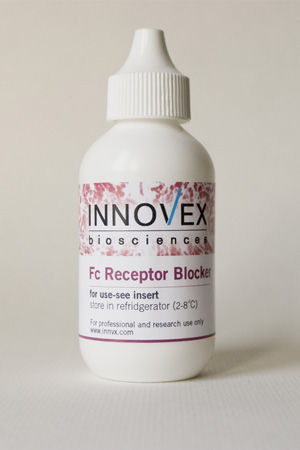
Innovex Fc Receptor Blocker is a UNIVERSAL Fc Blocker applicable to Blocking all types of Fc receptors such as Fcgamma receptors of type I, II and III; Fc-epsilon receptors type I and II; Fc–alpha receptors, Fcα/µR and FcRn. Innovex
Fc Blocker does NOT contain antibodies, Immunoglobulins or immunoglobulin fragments. INNOVEX Fc Blocker can be
universally used to block all types of Fc receptors for all-species’ cells including human, mouse and all-animal species cells and
tissues by a variety of Immunoassays such as IHC, Immunofluorescence (IF) and Flow cytometry. The Fc Blocker is also commonly used in eliminating background staining in Brain cells/tissues.
Fc Receptor blocker is also used for obtaining specific staining for tissues stained for kappa, lambda antibodies and
Immunoglobulins (lgs) by IHC, IF and Flow cytometry assays.
Format: Ready to use; no dilution or adjustments required.
Storage: Store in refrigerator at 2-8
o.C through the expiration date noted on the vial label.
Instructions for use:
For Blocking Fc receptors for IHC Staining:
1. Deparaffinize paraffin section slides OR cut frozen sections, fix and rinse in water as usual.
2. Quench endogenous peroxidase by immersion in 3% H2O2 (only for Peroxidase-IHC staining).
3. Cover sections or smears with 3-6 drops of Fc receptor block to achieve full specimen coverage.
4. Incubate for 30-minutes at room temperature.
5. Rinse with rinse buffer.
6. Proceed with the remainder of IHC staining steps per lab protocol.
The Fc Receptor Blocker must be used prior to use of any other blocker, e.g., serum or protein blocks.
The Blocker can be used in autostainers as a pre-treatment step prior to application of protein and/or
serum blocking.
For Immunofluorescence labeling of tissues and cell preparations:
Deparaffinize paraffin section slides OR cut frozen sections, fix and rinse in water as usual.
1. Cover tissue sections or cell preps with 3-6 drops of Fc Receptor Blocker to achieve full specimen coverage.
2. Incubate for 30-minutes at room temperature.
3. Rinse with PBS or DI water and proceed with application of fluorochrome-conjugated antibody (direct method) OR
with the application of non-conjugated primary antibody followed by fluorochrome conjugated secondary antibody
(indirect method).
The Fc Receptor Blocker must be used prior to use of any other blocker, e.g., serum or protein block or IF Blockers.
For Flow Cytometry & Blocking of Fc receptors:
1. Lyse or ficol blood as usual OR use whole blood
2. Add 150 to 300 microliter of Fc receptor block for 106
(million) cells.
3. Incubate for 30-minutes on ice OR at room temperature.
4. Wash twice in assay wash buffer.
5. Proceed with antibody labeling.
*This is a Research Use Only product.
